AI and glaucoma: a project exploring image analysis
A neural-network project for eye-image analysis: how the data is prepared and the limits of its use as clinical support.
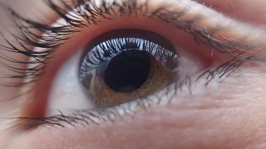
Glaucoma can damage the optic nerve and cause vision loss. The WHO’s global vision-impairment figure is not specific to glaucoma: at least 2.2 billion people have near or distance vision impairment, and in at least 1 billion cases it could have been prevented or remains unaddressed.
Editorial correction, 25 October 2026: the units in the original figure and its attribution to glaucoma have been corrected. The project retains its original 2022 publication context.
This article describes a research proposal using eye images and neural networks to explore support for clinical analysis. Results on one dataset do not establish a diagnosis or demonstrate that a tool works in other settings or populations.
How were the images processed?
Fundus images were used to train the neural network. After review by specialists, they were organised into two groups:
- Suspected glaucoma
- Patients considered healthy
The total number of images was also recorded; it is not a third class.
Defective images were removed before training the model.
Jupyter Notebook
In the case of the study carried out at the Salesian Polytechnic University, it was used Python in Jupyter Notebook , an environment for running and documenting Python code. It is open-source software that requires an installed environment and its dependencies. Images were organised into folders for healthy patients and others with suspected glaucoma.
Images in the study were cropped to isolate the optic disc, the region of interest. The cropping algorithm had two aims: first, to produce an image normalised to an approximately square shape and second, to keep the region of interest centred.
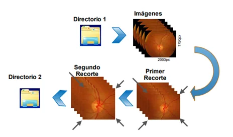
Algorithm used to crop images.
Image sizes were then standardised. Some images were used to train the classifier, while others were reserved for evaluating its predictions with a confusion matrix.
Search for optimal solutions
The proposal shows how to prepare images and train a classifier. Assessing its clinical usefulness would require independent evaluation, representative data and review by healthcare professionals. Performance depends on data quality, model design and how training and test sets are separated; high accuracy alone does not establish clinical safety.
If you want to be part of the search for optimal solutions, do not hesitate to start or continue your training in Artificial Intelligence with us. Check out the Saturdays.AI courses and choose the one you like the most in our website.
Bibliography
1.-Buri Abad, Edison Arturo. Processing digital images of the fundus with the use of artificial intelligence to provide a support tool for the presumptive diagnosis of human glaucoma. Feb-2022.
2.-Mayo Clinic. Glaucoma. Consult at: https://www.mayoclinic.org/es-es/diseases-conditions/glaucoma/symptoms-causes/syc-20372839#:~:text=El%20glaucoma%20es%20un%20grupo,personas%20mayores%20de%2060%20a%C3%B1os.
Written by
Eva Arnall

Saturdays.AI
Saturdays.AI is an impact-focused organization on a mission to empower diverse individuals to learn Artificial Intelligence in a collaborative and project-based way, beyond the conventional path of traditional education
A story from the Saturdays.AI community. Originally published on May 2, 2022.
← More community stories