Transformation of Angiotomography Images through the use of Artificial Intelligence.
Latam online. Second Edition. 2021 Contrast angiotomography What is the problem? The challenge Intravenous contrast media used in contrast angiotomography can have an impact on health...
A learning project shared by the community. The article documents its team, methods and results; it does not establish that the prototype is ready for real-world use.
Editorial note: this article documents an educational project and its results on the data used. It does not establish clinical validation or authorisation to diagnose or treat patients. Proposed healthcare applications are retained as the team’s goals, not demonstrated capabilities.
Latam online. Second Edition. 2021
Contrast angiotomography What is the problem?
The challenge
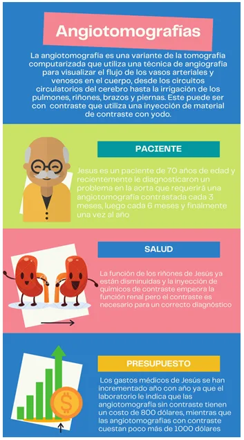
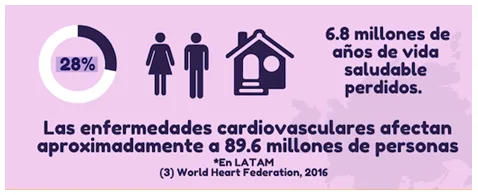
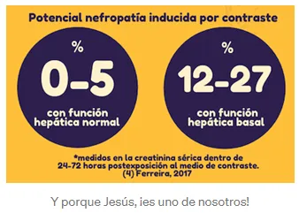
The intravenous contrast media used in contrast-enhanced angiotomography can have an impact on the nephrological health of patients, especially in those who have a history of high blood pressure, hypercholesterolemia, genetic history, older adults (60 years and older) and an increased cardiovascular risk (1), that is, they are more prone to:
- have strokes,
- acute myocardial infarction,
- peripheral arterial disease and,
- diseases of the aorta in general
There is the possibility of performing simple angiotomography that does not require the use of contrast media and has a lower cost (around $200.00 difference), but these do not allow complete visualization of the aortic structures clearly (2), so they are not useful in many cases, such as that of Jesús.

Why did we choose Angiotomography?
The opportunity. How do we propose to solve it?
ANGIOP.AI (CT Angiography Image Transformation System) explores AI-based medical image translation. The team aims to investigate whether, in future and after appropriate clinical validation, this technique might reduce the need for some contrast-enhanced studies. The prototype does not yet demonstrate that benefit.
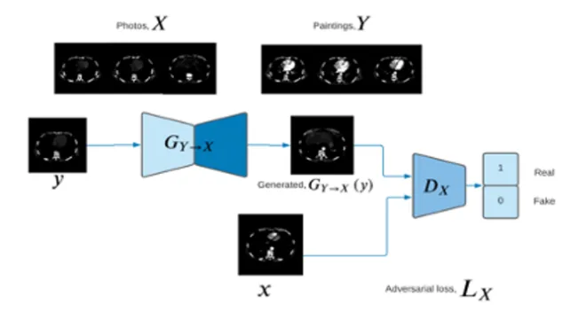
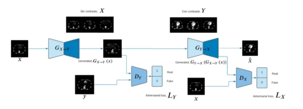
Figure 1. Structure of the GAN model applied to the transformation of Angiotomography images
ANGIOP.AI uses CycleGAN to transform non-contrast images into synthetic images resembling contrast-enhanced scans. The model learns to map images from domain X (without contrast) to domain Y (with contrast) without requiring exact pairs. A realistic-looking image does not guarantee preservation of clinical information.
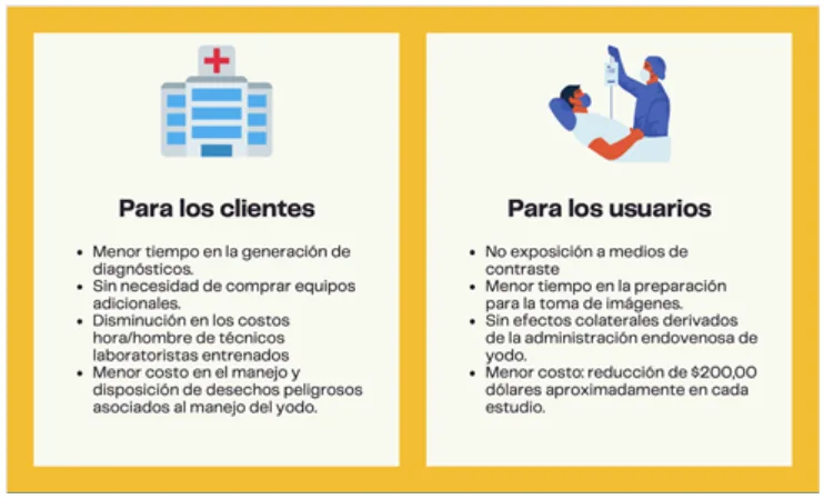
What are the benefits?
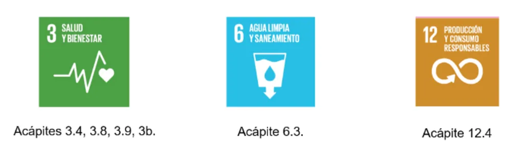
ANGIOP.AI is aligned with the fulfillment of the United Nations sustainable development goals for 2030.
We explain it step by step: methodology, model used and datasets
The datasets used correspond to those provided by Dr. Gonzalo Pullas, director of the Medicine degree at the University of the Armed Forces; who provided the images of angiotomography with contrast and simple angiotomography (without contrast) of 10 anonymous patients.
The team received 5,144 grayscale images measuring 512 × 512 pixels. Exploratory analysis examined formats, possible outliers and differences in pixel distributions across regions and slices. The images covered the same anatomical region but were taken at different times: they were not exact one-to-one pairs.
The model used is CycleGan with -1 to 1 scale image pre-processing for model input. The discriminators are convolutional networks with 5 layers that receive 256x256 images. The 4 layers are pattern definition and a classification layer. The generator uses three convolutional layers and six residual blocks. To calculate the cost functions, both for real and fake images, the mean squared error process is being used.
The model uses the following parameters:
- 20000 epochs
- Learning rate of 0.0001
The following steps were applied for training:
- Select a number of real images
- The generator takes the real images and adds noise to them to create a number of fake images
- Train the discriminator, making it classify the images as false or true a certain number of times or epochs
- Generate another number of fake images to train the generator
- The model is trained
- Review loss curves and generated images to assess training. This technical inspection does not replace clinical validation.
Let's see the results:
Lessons learned
- Teach the model to validate the input images if they correspond to the angiothoracic area.
- Increase the Dataset for future training.
- Validation of the generated images with a panel of medical experts is required.
What's coming: Future of ANGIOP.AI
- Use the data generated to measure the impact of the use of the system on reducing the incidence of kidney diseases.
- Develop a Web application.
References
(1) Cueva Torres, Dr., F. (2021). Epidemiology and Management of Aortic Diseases — SIAC. Inter-American Society of Cardiology.
(2) RadiologyInfo for patients (2020). Contrast Materials.
(3) World Heart Federation (2016) World Congress of cardiology & Cardiovascular Health
(4) Ferreira, J (2017) News in contrast nephropathy. Pontifical Bolivarian University, Medellín—Colombia. ELSEVIER Volume 14 Number 2
Members
Diego Chiza, Ana Gayosso, Gabriela Jiménez, Paola Peralta, Patricia Roman José Daniel Sacoto, María Teresa Vergara, Hilario Villamar, David Medrano.
Project presentation: DemoDay
Repository
The source code for this project can be found at: GitHub
More artificial intelligence!
Saturdays.AI makes AI more accessible through shared learning and projects with a social purpose. Our motto is AI4ALL IS AI4GOOD.
A story from the Saturdays.AI community. Originally published on March 9, 2022.
← More community stories